The Anatomy of the Femoral Vein
A Large Paired Vein of the Thigh
MedicalRF.com/Getty Images
Medically reviewed by Scott Sundick, MD
A large blood vessel of the thigh, the femoral vein is a major pathway in which blood from the lower limbs travels on its way back to the heart. It is sometimes called the superficial femoral vein (in contrast with the deep femoral vein).
This paired vessel, meaning it is located in both legs, is the main deep vein of the thigh, making it critical for lower limb and foot function.
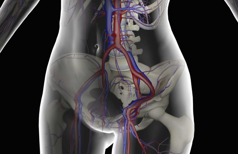
The popliteal vein, located behind the knee and deriving blood from the two tibial veins in the lower leg, continues into the femoral vein in the thigh, which then continues into the common femoral vein in the upper thigh. From there, blood passes through the external iliac vein in the pelvis.
Given its location and function, doctors may use the upper portion of the femoral vein for catheterization to diagnose and treat some cardiovascular conditions. Clotting of the femoral vein—a condition called deep vein thrombosis (DVT)—can lead to significant symptoms as well as pulmonary embolism (a clot in the lungs) should the clot break free and travel to the lungs.

MedicalRF.com / Getty Images
Anatomy
Structure
Among the body’s larger vessels, the diameter of the femoral vein in adults ranges from approximately 12 millimeters to 14 millimeters (mm), which is about half an inch. Emerging as it does near the knee, it tends to grow in size as it moves up the thigh, with its wider regions being closer to its terminus, near the groin.
Like all vessels in the body, the femoral vein is made up of three layers of cells:
Tunica intima is the inner lining of the vein. It’s composed of squamous epithelium, a semipermeable layer of cells, as well as connective tissue.
Tunica media is a relatively thick middle layer, composed of smooth muscle, which can apply pressure to help push blood along.
Tunica extrema, the outer lining, is composed of varying amounts of elastic and rigid fibers. These shape the vein and help keep it in place.
Location
As mentioned, the femoral vein forms after the popliteal vein runs up the back of the knee and passes into the adductor hiatus, which is an opening between the adductor magnus muscle of the inner thigh and the femur.
It then passes through the anterior aspect (front of) the thigh, running upwards and towards the center of the body along a groove called the adductor canal. Along this course, it accesses the femoral triangle, a depression between the thigh muscles, where the femoral vein runs next to the femoral artery, a major supplier of blood to the lower limbs.
It crosses the femoral sheath, a funnel-shaped space connecting the lower abdomen and femoral triangle. Then the femoral vein terminates and turns into the external iliac vein, just behind the inguinal ligament, a tough band of tissue that forms a barrier between the thigh and the pelvis. In turn, blood drains into the common iliac vein and eventually the heart.
There are several important tributaries that drain into the femoral vein as it moves upward and medially (towards the center of the body) through the thigh. They are the following:
Deep femoral vein: This vessel, the other major vein of the deep thigh, accesses the rear of the femoral vein about 8 centimeters (a little over 3 inches) from the inguinal ligament.
Great saphenous vein: A large superficial vein on the inside of the leg, this vessel runs from the foot to the thigh through the subcutaneous tissue below the skin of the lower leg. It joins the femoral vein on its front side, close to the pelvis.
Circumflex femoral veins: The lateral and medial circumflex femoral veins connect to corresponding circumflex femoral arteries, which are branches of the deep femoral artery of the leg.
Anatomical Variations
Generally speaking, congenital anatomical variations of the femoral vein are relatively common. The most common of these include:
Duplicated femoral vein is the most common abnormality, in which a second, parallel femoral vein runs alongside the original.
Axo femoral trunk is a case in which the femoral vein does not fully form, making the axial vein the primary vein of the thigh.
Deep femoral trunk occurs when lack of development of the femoral vein causes the deep femoral vein to be the primary pathway for blood leaving the lower limbs.
In many cases, too, doctors will find differences between the structure of the veins in the left and right thighs.
Function
Veins take deoxygenated blood (blood depleted of oxygen after being absorbed by the cells) back to the right side of the heart. The right side of the heart then carries this blood to the lungs so the blood can become oxygenated. The oxygenated blood then travels to the left side of the heart, and the left side of the heart pumps the oxygenated blood out to the body.
As the primary deep vein of the leg, the femoral vein is critical for draining blood from the lower limb.
Clinical Significance
Because of its larger size, in the hospital and healthcare setting, the femoral vein may be used in a couple of important procedures, and it may be implicated in some health conditions. Here’s a quick breakdown:
Catheterization
In this procedure, a small tube is run through the femoral vein to access the right atrium of the heart, where it can measure blood pressure and oxygen levels. Catheterization is most often used to diagnose and treat certain cardiovascular conditions, such as heart failure and coronary artery disease.
Venous Sampling
Used as a means to collect samples of vein tissue for testing and assessment, this procedure helps doctors diagnose certain hormonal conditions or diseases, such as Cushing’s syndrome, aldosteronism (a type of high blood pressure), and hyperthyroidism, among others. The femoral vein can serve as an access point for tools tasked with collecting this tissue.
Deep Vein Thrombosis
DVT is a potentially dangerous condition in which a blood clot forms in a deep vein. Often occurring in lower limb veins like the femoral vein, DVT becomes especially serious if clotted material breaks off and reaches the lung (a condition called pulmonary embolism).
Not only does this lead to swelling, pain, and tenderness, if it pulmonary embolism occurs, it can become a life threatening emergency.
Related: What Is Deep Vein Thrombosis?

